Guide
Why the cone-beam CT matters
A flat X-ray cannot show bone thickness or where the nerve runs. Here is what a cone-beam CT does, and how to check whether a clinic has one.
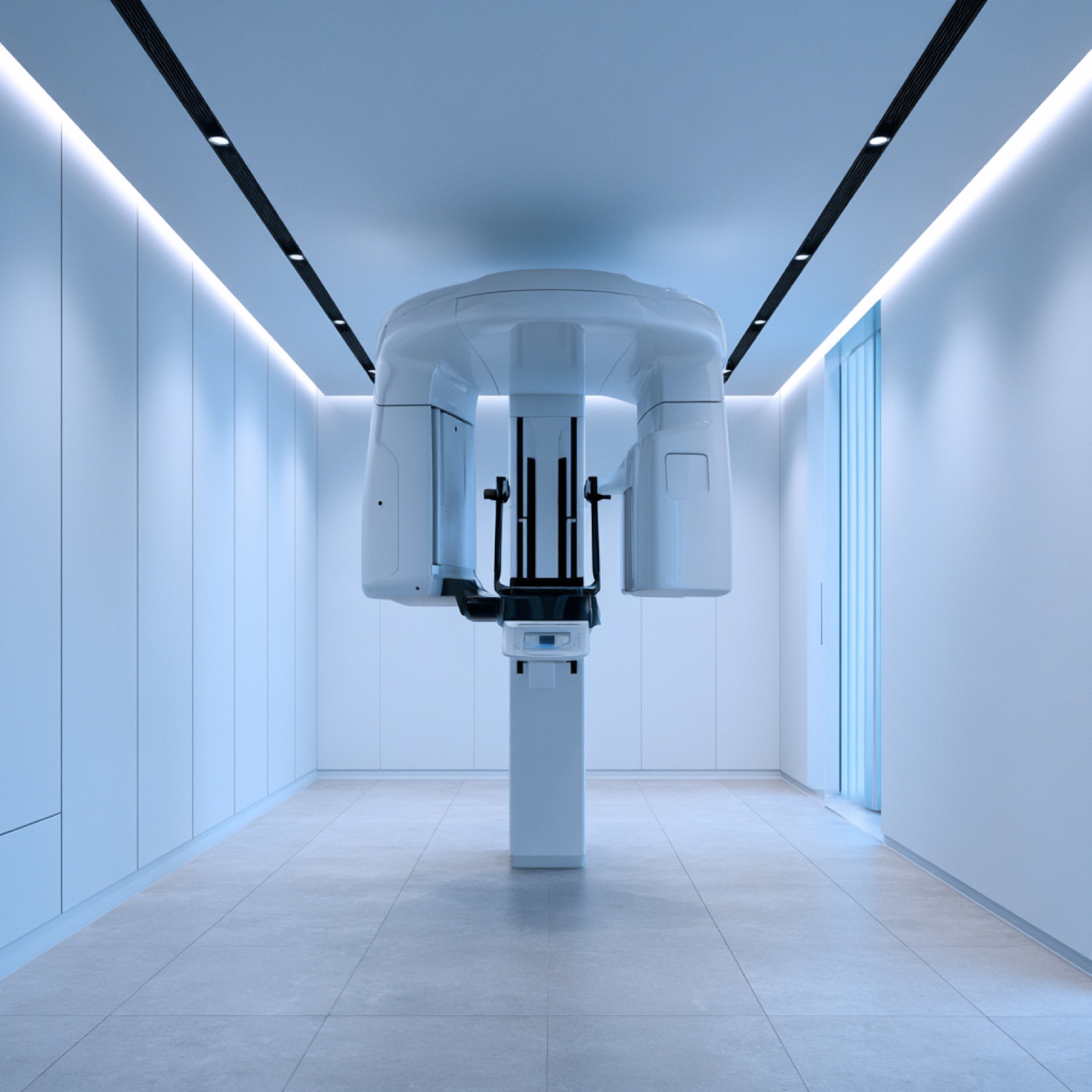
Panoramic X-ray versus cone-beam CT
The panoramic X-ray most dental visits start with is a two-dimensional image of the whole mouth unrolled onto one sheet. It is good for scanning the arrangement of teeth and spotting larger lesions, but it cannot show the thickness of bone or the distance to the nerve canal. On a map, it is the plan view with no cross-section.
A cone-beam CT (CBCT) images in three dimensions. It shows bone width, height and density, and the position of the inferior alveolar nerve canal and the maxillary sinus. Planning where an implant goes — at what depth and angle — needs that information.
What it changes
With a cone-beam CT, the need for a bone graft is judged from evidence rather than impression, and risks such as nerve injury can be designed around at the planning stage. "We can place one" with no imaging and "the bone here is this many millimetres wide, so" with a cross-section on screen are not the same consultation.
Orthodontics uses CBCT too — to locate an impacted tooth, for instance. It is not required for every orthodontic case; whether to image depends on what the diagnosis needs.
Whether a clinic has one is public record
A cone-beam CT is equipment clinics register with HIRA, so which clinic has how many is verifiable from open data. The area comparison pages on this site print the registered count for every clinic exactly as filed.
More units does not mean better diagnosis. But if an implant plan arrives without any cone-beam CT, it is fair grounds to ask what the plan was read from.
FAQ
Frequently asked
Does the scan hurt, or carry risk?
It takes a few dozen seconds, standing or seated, and does not hurt. The radiation dose is lower than a medical CT, but tell the clinic before imaging if you are pregnant.
Is a cone-beam CT mandatory before an implant?
It is the standard record for reading bone condition and nerve position in three dimensions. If a placement plan appears without one, it is reasonable to ask what it was based on.
What does the scan cost?
It varies by clinic and by scan area, and is generally not covered by insurance. Ask about the imaging fee when you book, and whether it is deducted if you go ahead with treatment.
Is a panoramic X-ray not enough?
A panoramic is a two-dimensional overview; it cannot tell you how thick the bone is or how far the nerve sits. Useful for a first check-up, not sufficient for planning placement.
What is different about a clinic with two or more units?
A practice large enough to run several chairs or floors may simply have shorter waits. Diagnostic quality itself rests on how the images are read and planned from, not on how many machines there are.
Can I take my scan data with me?
It is your own medical record, so you can request it on a disc or as files (a small issuing fee may apply). It is what saves you a repeat scan when consulting elsewhere.
Is a cone-beam CT needed for orthodontics?
Not as a rule. It is taken when the diagnosis calls for it — locating an impacted tooth, checking root condition. The standard orthodontic records are a lateral cephalometric X-ray and a scan.
What is guided or "navigation" implant surgery?
Planning the placement from cone-beam CT data and fabricating a surgical guide to follow. Clinics use different names and apply it to different extents, so ask how much of the process is done digitally.
Related